Complete AI Stroke Imaging

69edf8e12792d2380d591e6b_Logo

Brainomix 360 Stroke

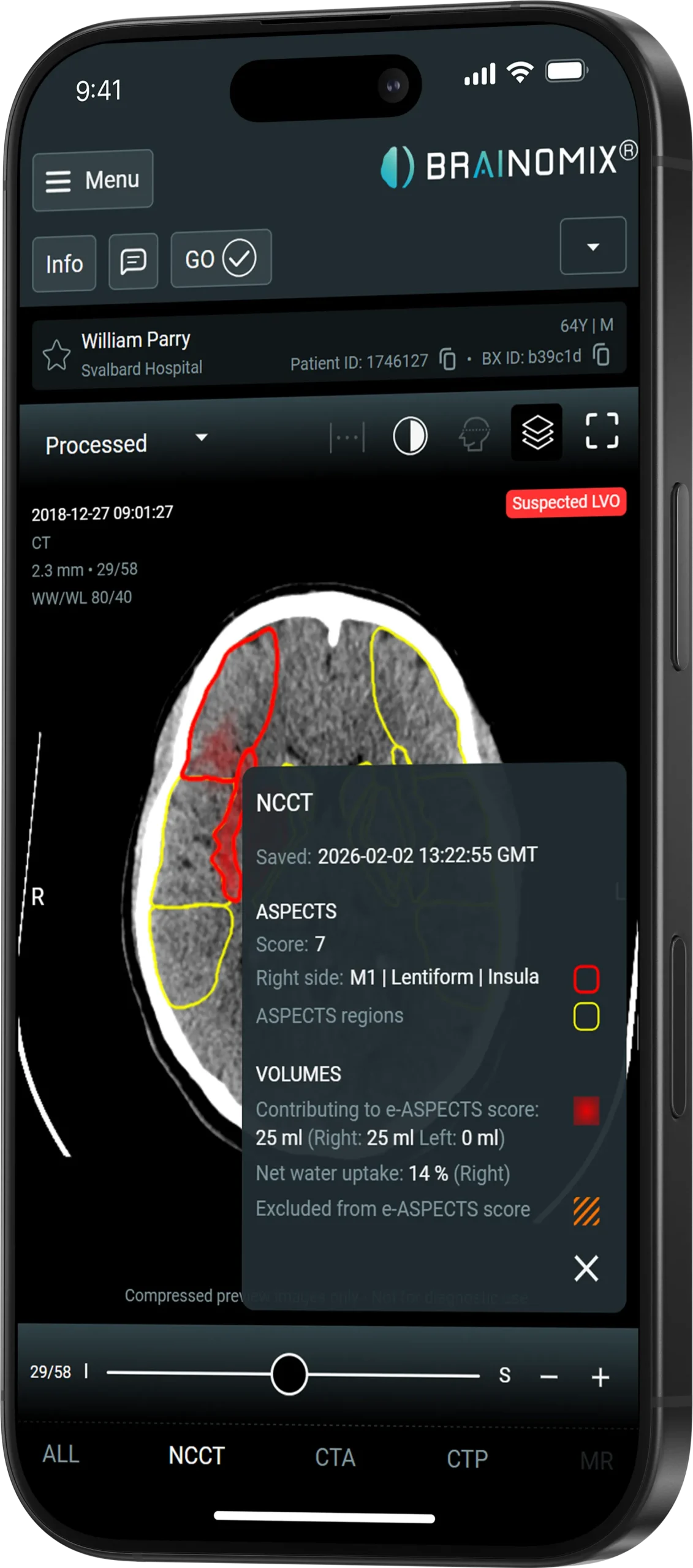
Brainomix NCCT suspected LVO and intracranial hemorrhage analysis

LVO + ICH Detection

Non-Contrast CT

Expert insight from routine CT.

Brainomix 360 NCCT analyzes standard non-contrast CT imaging to support faster assessment of hemorrhage, early ischemic change and suspected vessel occlusion.

Automated processing Results within minutes PACS + mobile delivery
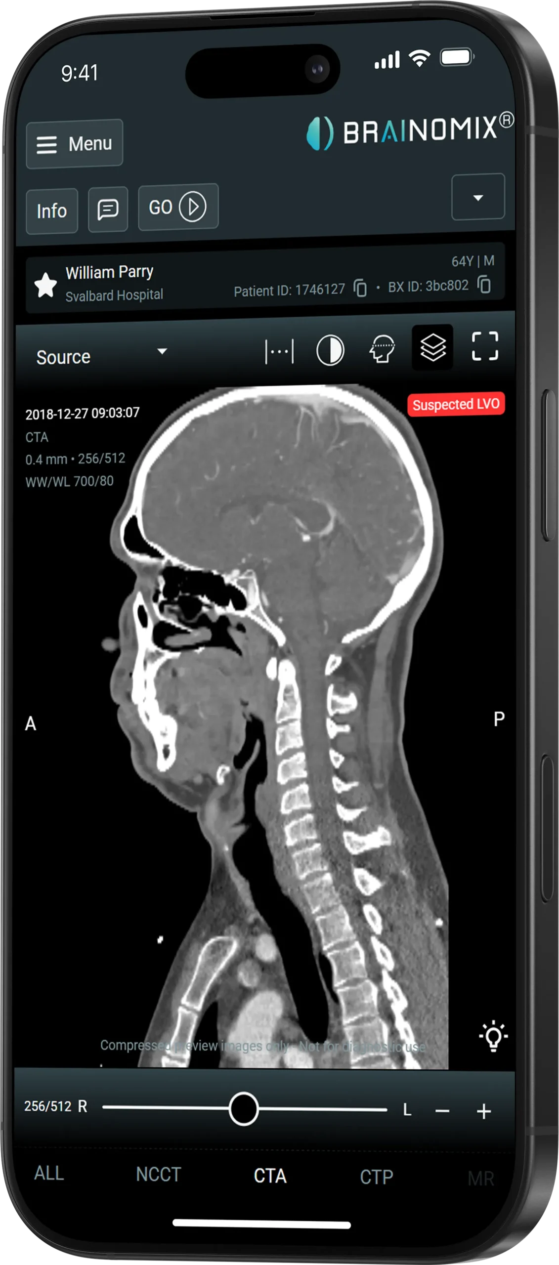
CT Angiography

See the vessel pathway with greater clarity.

Brainomix 360 CTA automatically detects suspected large vessel occlusions and delivers objective vessel-density information for treatment planning.
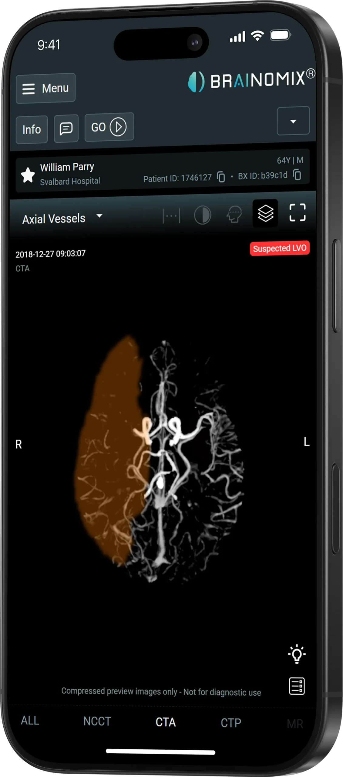
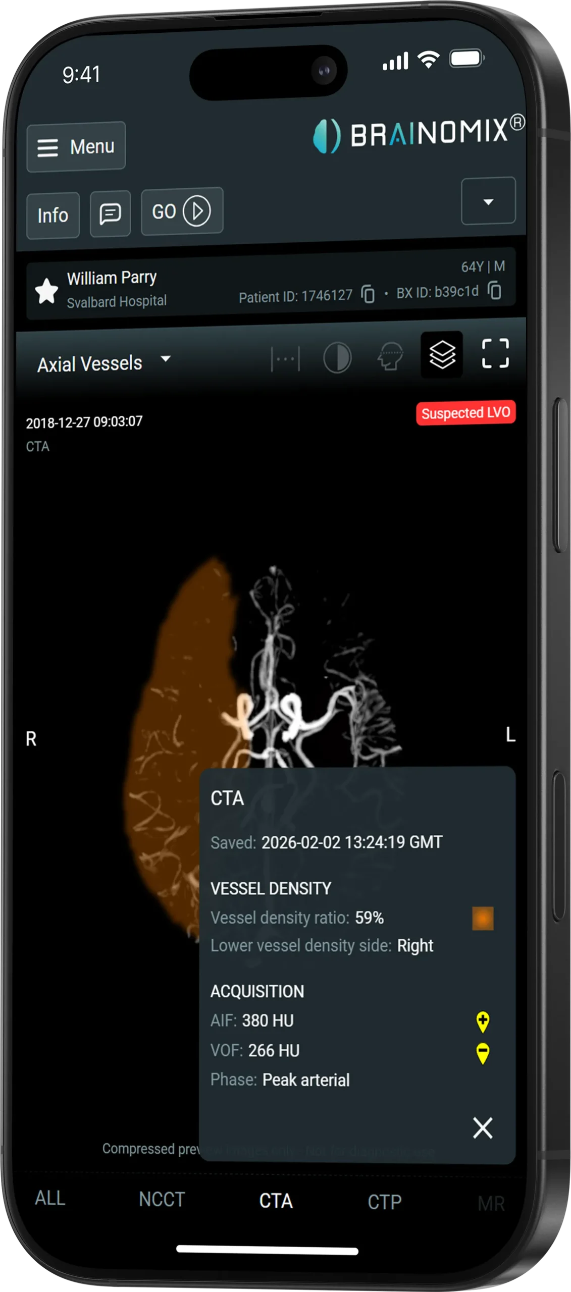
CTA large vessel occlusion detection
CTA vessel density heatmap and scoring
CTA multi-planar reconstructions
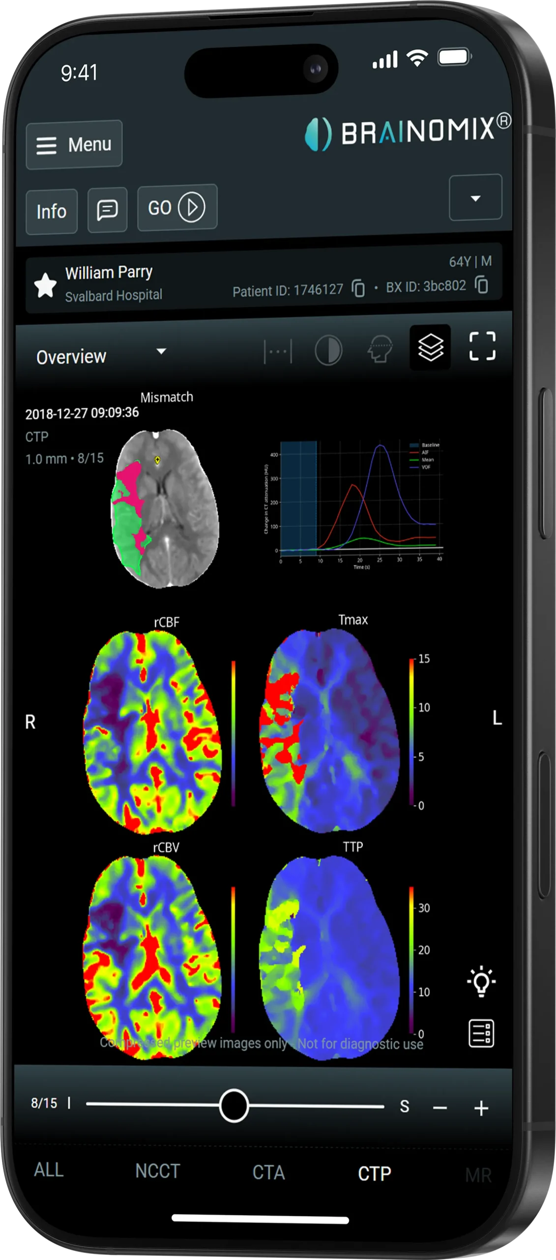
CT perfusion multi-parameter maps
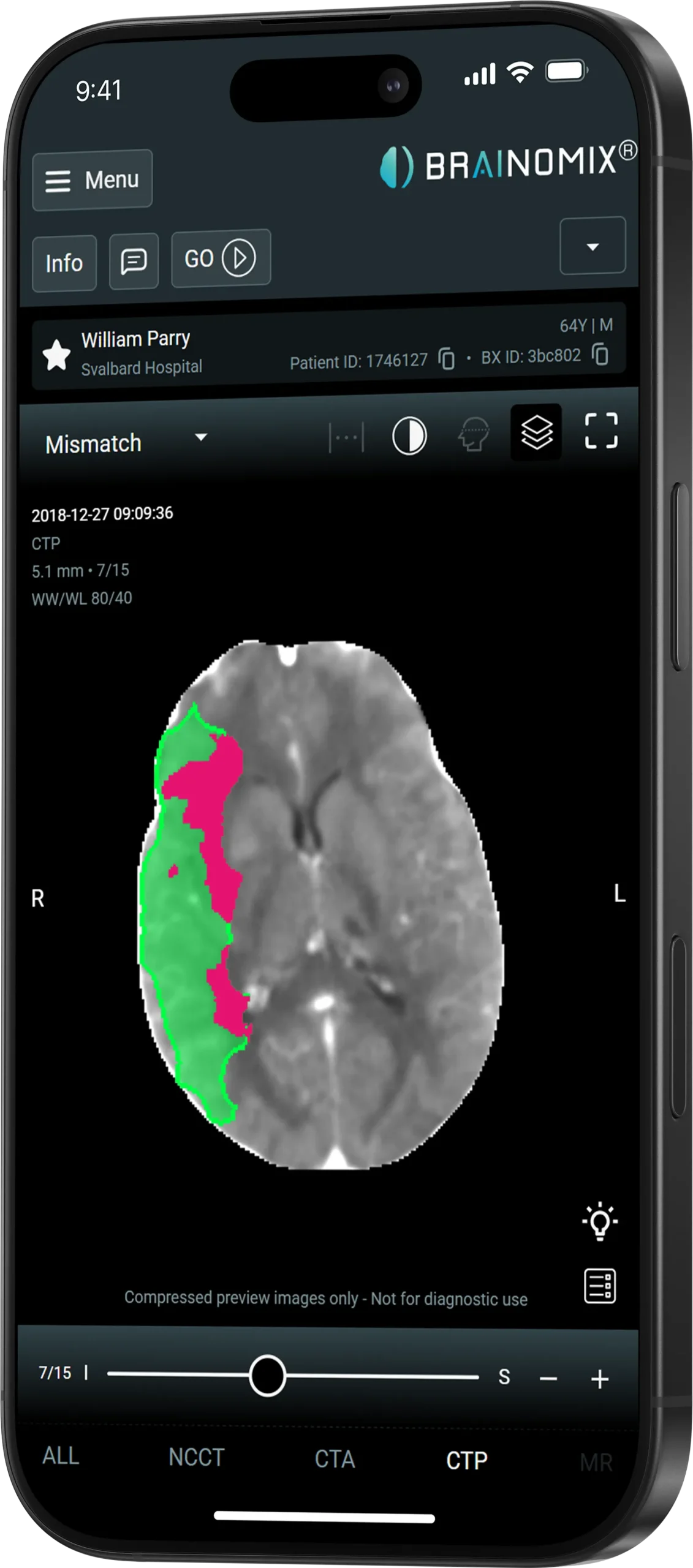
CT perfusion automated mismatch analysis
CT perfusion core and Tmax volume analysis
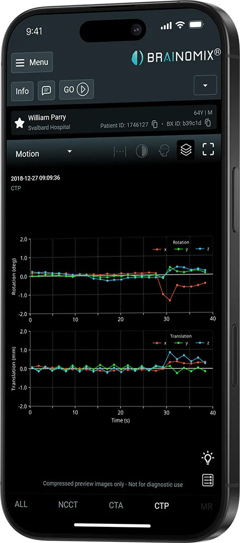
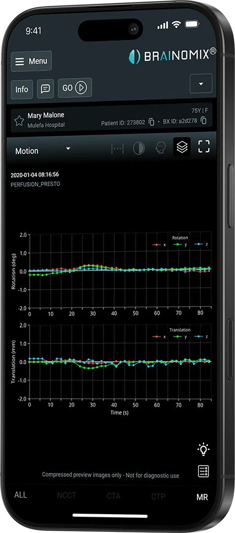
CT perfusion motion correction analysis
CT Perfusion

Clear perfusion insights.

Brainomix 360 CTP transforms perfusion imaging into visual maps and quantitative results, helping teams assess salvageable brain tissue.

Quality Maps Core + penumbra Tmax + rCBF EVT assessment support
Magnetic Resonance Imaging

Advanced MRI insights.

Brainomix 360 MRI brings diffusion and perfusion information into one connected workflow, supporting faster and more informed tissue assessment.

MRI automated mismatch map
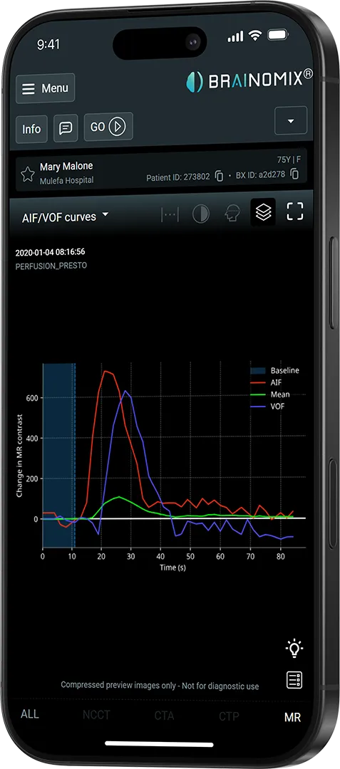
MRI motion correction analysis
MRI multi-parameter maps
Brainomix 360 Stroke

See Brainomix 360 Stroke in action.

Discover how AI-powered imaging can support faster, more informed stroke treatment decisions.

Request a demo

Product Enquiry