Lesion Quantification
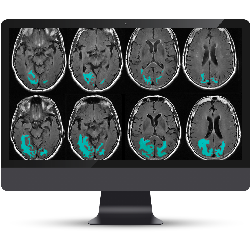
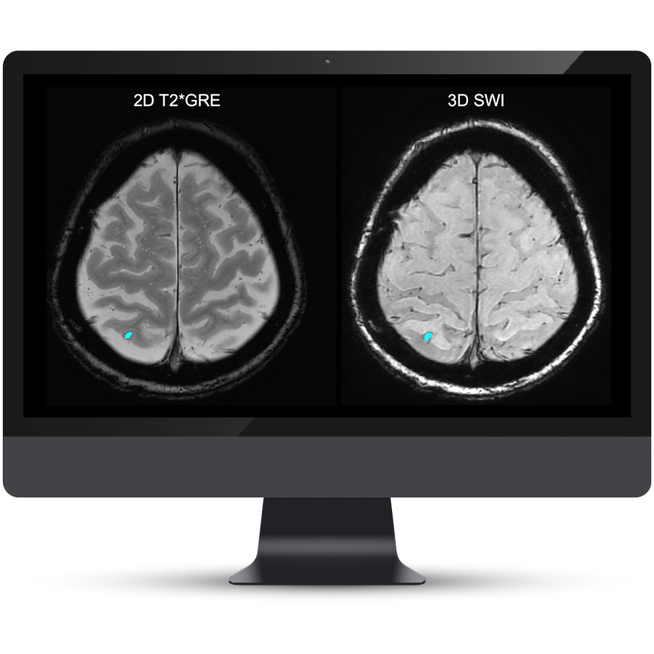
Accurate segmentation and longitudinal tracking of FLAIR, T2* GRE, and SWI lesions.

Automated brain lesion quantification, visualization, and tracking over time to support clinical assessment, including monitoring patients receiving anti-amyloid therapy.
NeuroQuant® Lesion Surveillance quantifies brain lesions and tracks changes over time, supporting radiologists and neurologists in assessing and monitoring brain changes, including those that may occur in patients with Alzheimer’s disease receiving anti-amyloid therapy. Consistent, structured measurements across baseline and follow-up MRI scans help clinicians assess treatment eligibility and monitor therapy-associated imaging findings.
Support clinical assessment with objective volumetric data and color-coded brain structure images to inform diagnosis, treatment, and lifestyle planning. Assess lesion dissemination and distribution patterns, identify areas with the greatest lesion burden, and track lesions across orthogonal views.
NeuroQuant®
Accurate segmentation and longitudinal tracking of FLAIR, T2* GRE, and SWI lesions.
Objective lesion quantification at baseline, helping clinicians document and contextualize findings.
To ensure reproducible outcomes and accuracy over time.
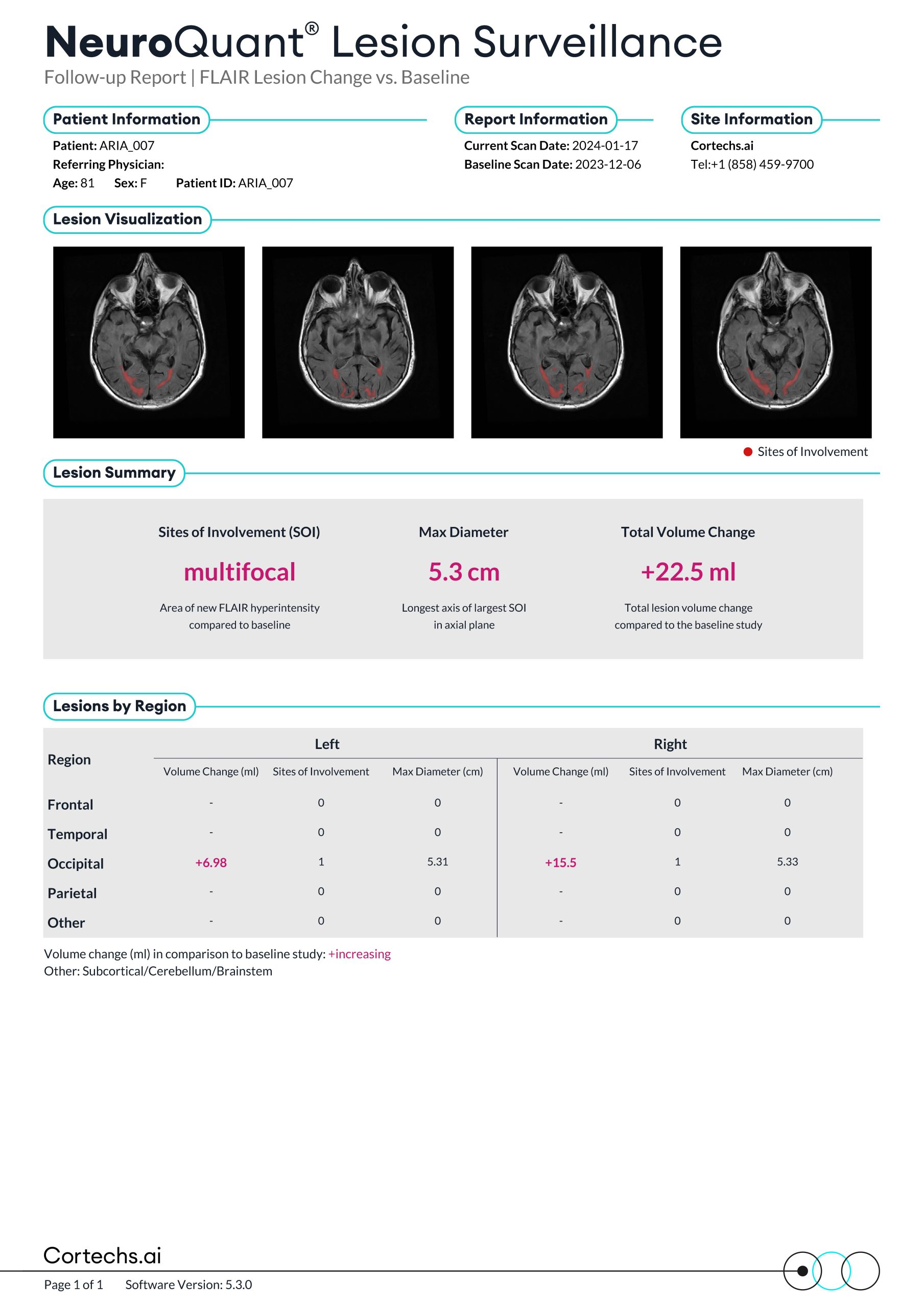
Monitor lesion counts and maximum diameters over time, with comparisons to baseline and prior timepoints.
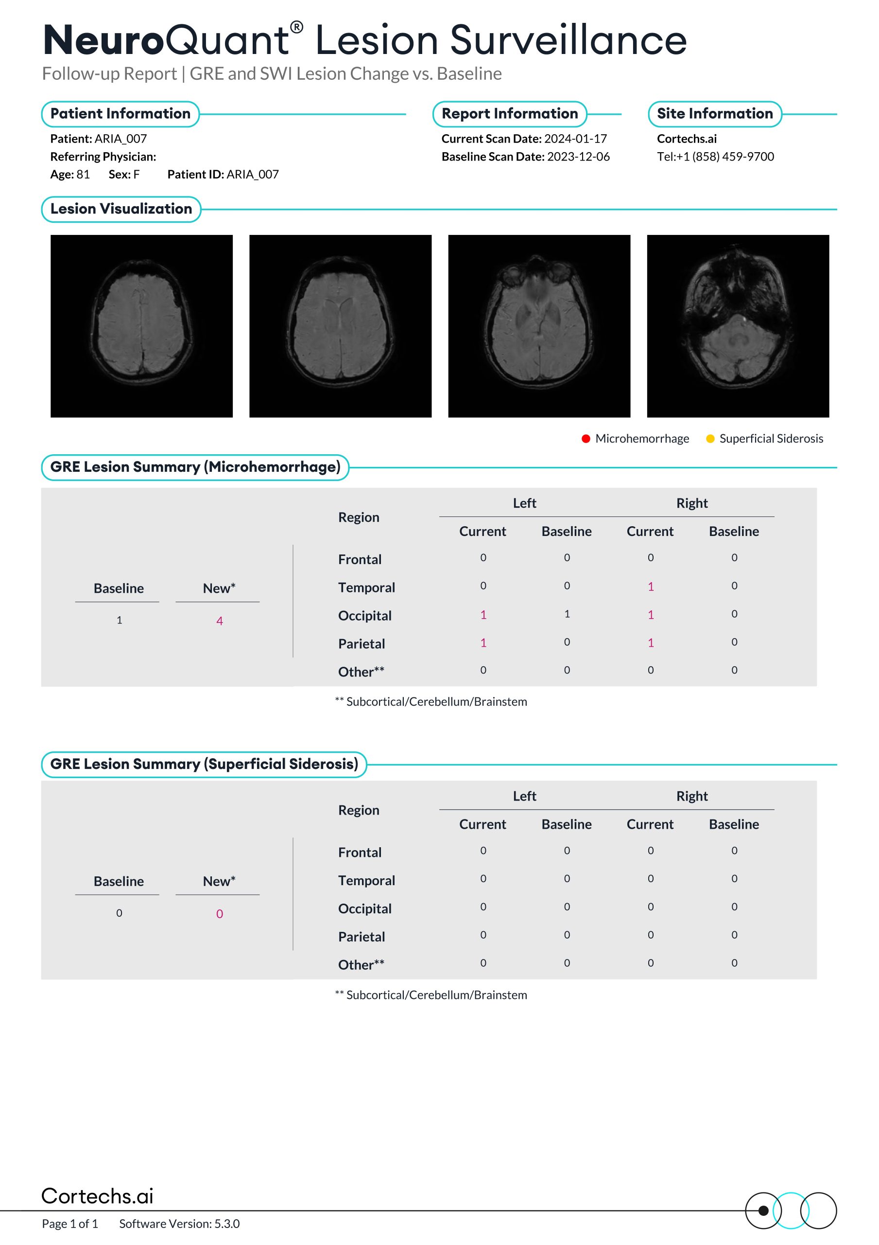
Quantitative reports that support physicians in monitoring FLAIR and T2* GRE/SWI lesions over time through comparison with baseline imaging.
The NeuroQuant® Lesion Surveillance GRE Report analyzes and tracks T2* GRE/SWI lesions, providing counts and locations of microhemorrhages and superficial siderosis.
Take the next step
NeuroQuant® Brain Tumor
Powered by AI, NeuroQuant® Brain Tumor assists radiologists, oncologists, neurosurgeons, and the Tumor Board by providing objective quantification and analysis of tumor changes over time.
Request a demoLet’s discuss your brain tumor imaging workflow.